Thursday, July 7, 2011

Treatment of Periodontitis

Periodontitis - an inflammation of the periodontium (periodontal tissue collection, which provide connection to the root of the tooth with hole). Periodontitis is a common and very serious illness. In fact, periodontitis - is running periodontitis (gum disease).


Periodontitis is treated quite successfully in any dental clinic. But periodontitis completely fails to cure, as the disease is chronic. Delays in treatment of periodontitis or any other form of gum disease, usually results in the final result in tooth loss.

In addition, periodontitis may be associated with reduced immunity, give rise to diseases of the gastrointestinal tract.

Treatment of periodontitis

In the dental clinics in Moscow offers a comprehensive approach to the treatment of periodontitis with the obligatory account the individual characteristics of the patient.

What we need is a thorough examination of the mouth. Diagnosis is based including the X-ray study methods, which include proper intraoral x-ray, CT scan and orhtopantomogram. These measures help determine the severity of the disease.

Also, a thorough explanation of somatic disease patient, living habits and the presence of hereditary factors. In the treatment of periodontitis in the dental widely used method of ultrasonic scaler, laser therapy and photophoresis - the introduction of a laser beam to the gums of drugs.

In particularly difficult cases, specialists perform surgeries to help decrease tooth mobility. It is also possible "pulling" the gums, bone grafting and the use of special membranes. After elimination of the inflammatory process, patients are offered at special techniques needed prosthetics to preserve the integrity of the dentition.
Combined treatment of periodontal disease is impossible without total body strengthening and enhancing immunity.

Prevention of periodontitis

To achieve a positive effect of treatment of periodontitis and store the result in the long term is very important to the prevention and elimination of chronic disease patients.  Along with professional treatment is necessary to conduct their own preventive measures at home. First, the patient requires discipline. Regular and thorough oral hygiene is an integral part of prevention of periodontitis.

On the recommendation of a physician can rinse your mouth daily green tea or infusion of oak bark (two tablespoons of bark pour a glass of boiling water, infused for thirty minutes and filtered). It is also useful after brushing rinse mouth chamomile extract, which is known to have antibacterial and wound-healing effect.

It is known that any disease is easier to prevent than to cure. Therefore, careful oral hygiene, keeping the overall tone of the body, treatment of chronic diseases, as well as regular visits to dental clinics should be the norm of modern life.

Tuesday, July 5, 2011

Children Periodontitis - Causes and Classification of Child Periodontitis

Children periodontitis
The structure of dental caries disease complications account for about 35-50% of all cases of requests for dental care. The number of caries complications, including periodontitis, is constantly high, even among the urban population of Russia. And worse than organized sanitation, the more frequently diagnosed with inflammatory periodontal disease.


Periodontium is located in the space bounded on one side of the cortical plate wells, on the other - the root of the cement. Feature of periodontal unformed tooth is that, extending from the neck of the tooth to form part of the root and merging with the growth zone, it is in contact with the pulp canal. As formation of the root zone of the size of the sprout and the apical foramen contact with the pulp are reduced, and periodontal ligament length increases. After the end of the root apex as far back as the year continues to develop periodontal. As the root resorption of temporary teeth periodontal ligament length decreases and the contact with the pulp and periodontal cancellous bone increases again.

During the period of temporary tooth root resorption at the site of its adjacency to the rudiment of constant cancellous and cortical plate, bounding the gap, and periodontal root cement, dissolve, which leads to the disappearance of periodontal at this site.

The absence of a stable structure and the thickness of the periodontium in the apical part is the anatomical and physiological features during the development and formation of the roots of temporary and permanent teeth.

Periodontium in the child presented a rather loose connective tissue and contains a large number of cellular elements and blood vessels, which accounts for its reactivity when exposed to adverse factors.

The primary cause of periodontal disease in children is when micro-organisms, their toxins, biogenic amines, coming from the inflamed necrotic pulp are distributed in the periodontium.

Second place goes to the tooth trauma: injury, dislocation, fracture of the root at some level. In this case we are talking mainly about the front teeth. Young children, toddlers, fall face down, resulting in different types of partial dislocations, including vkolochennyh, often accompanied by rupture of a neurovascular bundle. At school age, are often permanent injury immature anterior teeth. If children do not seek help, the pulp gradually, without marked clinical manifestations, dies, and develop chronic periodontitis. A role in periodontal disease may play a mechanical trauma during root canal treatment sharps, needles, or drilborami during injection of filling material over the top.

A role in the development of periodontitis in children are strong chemical and medicinal substances that penetrate into the periodontium in the treatment of pulpitis. Often there are cases of periodontitis, especially in the frontal teeth of the upper jaw, because of the reluctance to use insulating pads when working with evikrolom.
Reducing the time of treatment of patients with pulpitis by reducing length of stay of mummified in the oral cavity leads to the further progression of the disease process, the transition of a single nosologic entity to another. Was also found that 20% of the occurrence of periodontal disease was caused by the prolonged treatment of teeth with pulpitis, especially in cases plohoprohodimyh channels. In this case violated the rules of endodontic treatment channels, but since the method allows replacement of mummified devitalization of the antibiotics, enzymes, etc. In 4% of patients with periodontitis was caused by errors or complications during treatment: perforation of the bottom cavity of the tooth or root canal in the presence of endodontic instrument fragment, excessive excretion of filling material in zaverhushechnuyu area and 6% - incomplete root canal fillings. In 2% of patients with periodontal disease occurrence was associated with prolonged use of arsenic prepaoatov.

Thus, in 41% of cases due to the emergence of periodontitis iatrogenic factors. In rare cases, periodontal inflammation may develop hematogenous route with acute infectious diseases of children. Possible way of spreading the infection to the periodontium per continuitatem from inflamed tissues, in the neighborhood.
Features of the appearance and character of the course of inflammatory processes in the periodontium, but the direct etiologic factors are largely determined by local and also the general level of resistance. Such co-acute and chronic diseases such as flu, sore throat, gastrointestinal tract, etc., reduce the protective capacity and immune reactivity of the organism, creating conditions for the occurrence of acute and chronic periodontitis.

Of periodontitis exhibit different associations of microorganisms. In the composition of the microflora is dominated by grampolo of positive-negative cocci (mainly staphylococci and streptococci) and yeasts, lactobacilli, actinomycetes, etc. Among the most frequently isolated microorganisms in the first place - aerobic and anaerobic species of streptococci, and then - staphylococci.

In the diagnosis of chronic periodontitis temporary teeth in children can not be limited only by clinical data. Understanding of the nature of the inflammatory process and the extent of its influence on the permanent tooth bud give X-ray data. It must be done not only in the event of various complications of the inflammatory process, but before any treatment of a tooth with chronic periodontitis, as even during quiet clinical course of inflammation can be detected radiographically severe complications, changing treatment policy.

Of particular value in addressing this issue in pediatric practice of utilizing well-proven method of panoramic radiography. In a short time and at low doses of radiation it gives much information about the state of the periapical tissues of teeth, can identify the correct remedial measures to avoid complications, etc.

Classification of periodontitis
On the basis of isolated etiological infectious, traumatic and drug periodontitis. By location - apical and marginal. Downstream of the pathological process are acute (serous and purulent), and chronic (fibrosis, granulating, granulomatous), and exacerbated chronic periodontitis. Periodontitis is marginal, marginal, or if the inflammation is primarily originated from the gingival margin. The cause of his injury is often the gingival papillae in pencil, food, sharp-walled cavity, the edges of the crown. Prolonged irritation of the gums promotes the progression of acute inflammation in chronic marginal.

Acute apical periodontitis
The clinical picture of acute and pathoanatomical periodontitis temporary teeth is basically the same as permanent. However, the anatomical features of the temporary teeth are responsible for some originality in the development of acute periodontitis: in connection with a wide opening in the apical periodontium during formation or resorption of the roots of pulp inflammation is readily converted to the periapical tissue. Sometimes periodontitis develops earlier than the inflammation encompasses the entire root pulp. More severe periodontal disease and tooth temporary transition of the inflammatory process in the surrounding soft tissue and bone is associated with reduced resistance to the child's body.

In acute apical periodontitis children complain of pain expressed by the ever-increasing, with pressure growing on the tooth. The child indicates exactly a bad tooth. The next important symptoms are swelling of the gums and surrounding soft tissue swelling and an increase in regional lymph nodes, sharply painful percussion of the tooth. In temporary teeth acute periodontitis develops very rapidly and, if not created by the outflow of fluid, infection is spreading rapidly across the jaw bone, causing the abscess. In young children, in response to inflammation of periodontal growing general signs of intoxication: increased temperature, increased sedimentation rate and leukocytosis.
Acute apical periodontitis in children is rare, it must be differentiated from exacerbation of chronic periodontitis. The clinical picture of both diseases may be identical, but in acute periodontitis in the radiograph is no deviation from the norm, and an exacerbation of chronic periodontitis radiographically well-defined characteristic changes.

Prevention of acute periodontitis is an early, timely and proper rehabilitation of the teeth with pulpitis. Acute apical periodontitis can result in recovery and transition to the chronic form, and in case of increase of the inflammatory process - the development of periostitis or osteomyelitis.

Chronic periodontitis
In children, the inflammatory processes in the pulp and periodontium are closely linked. More than 30% of chronic periodontitis occurs when the incorrect treatment of teeth with pulpitis, about 38% - with the development of caries without treatment and about 30% - due to injury. Children 6-8 years chronic granulating periodontitis develops as a result of permanent injury immature incisor teeth (usually the top jaw) and due to decompensated acute course of caries in first molars. Chronic periodontitis develops as a result of acute apical periodontitis or chronic primary process as a result of chronic pulpitis. Picture in pathology of chronic periodontitis is dominated by the phenomenon of exudation are not as acute process, and proliferation: growth of fibrous or granulation tissue.

Chronic periodontitis in temporary and permanent teeth often immature in the presence of a shallow cavity in the absence of communication with the cavity of the tooth. This is due to anatomical features of the structure of hard tissues of temporary and permanent teeth with incomplete root formation.

Chronic periodontitis fibrotic

Complication arises as a result of pulpitis, periodontitis, acute and chronic treatment after granulating and granulite lematoznogo periodontitis. Develops only in teeth with formed roots (both temporary and permanent teeth). During the formation of the tooth, in the absence of a stable structure in the apex of the root, and root resorption during the temporary teeth fibrous periodontitis is not observed. Clinically, fibrous periodontitis is not accompanied by any symptoms and are sometimes found incidentally during dental X-ray system. There are no complaints, percussion painless, gum normal color of the tooth mobility is not detected. Sometimes the color of the tooth crown, he becomes more dull with a grayish or bluish tint.

Diagnosis is based on X-ray: fibrous thickening of periodontitis is characterized by periodontal and seal, as evidenced by extensive periodontal crevice. The width of periodontal ligament varies in a limited area or, less frequently, throughout the periodontal space, which depends on the extent of the inflammatory process.

Chronic periodontitis fibrous differentiate from secondary caries, chronic gangrenous pulpitis, to a lesser extent - from deep caries. Similar to chronic periodontitis fibrous X-ray pattern is observed at a time when the formation of the root apex over and over, the periodontal gap is physiologically enhanced. Increased periodontal ligament noted also in some types of partial dislocation of the stylus in the direction of the adjacent tooth. In this case, the gap narrowed periodontal on the side where the tooth was displaced, and extended from the opposite side of the displacement.

Chronic granulomatous periodontitis

As chronic fibrotic, granulomatous periodontitis in temporary teeth is rare. Develops mainly in permanent teeth with roots formed, asymptomatic, rarely formed a fistula in the area of the damaged tooth. Typically, cavities are not in communication with the cavity of the tooth. On X-ray revealed granulomatous periodontitis in the form of focal destruction of bone tissue round or oval at apex with clear contours up to 5 mm (Fig. 4). Bone tissue around the granulomas are usually not changed, at least on its edges visible sclerosal compacted zone delimited bone of normal structure. The presence of multiple sclerosis indicates the reaction of the bones with prolonged duration of the inflammatory process. Periodontal gap bounded by the cortical plate, there is not throughout the root. The tip of the tooth root, which is located in the granuloma, often not resorbed.

Differentiate chronic granulomatous periodontitis should be of secondary caries, chronic gangrenous pulpitis, and other forms of periodontitis, kistogranulemy, cysts and partial dislocation of the tooth towards the occlusal plane. Radiological case of incomplete dislocation in the direction of the occlusal plane is visible deserted part of the alveoli, resembling a granuloma. Some natural openings (copperplate, mental) are projected on top of the root, mimicking granuloma. In permanent teeth immature granuloma should be differentiated from the emerging growth areas of the tooth. Sprout area bounded continuous cortical plate. At granulite lematoznom periodontitis periodontal gap is uneven width, in the apical part of the root extended. Cortical bone can be traced only to that portion of the root, from which to begin the expansion of periodontal ligament.

Chronic granulating periodontitis
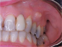
The most common periodontal inflammation temporary teeth becomes chronic granulating periodontitis. Most often the disease is asymptomatic in a shallow cavity, and this causes difficulties in diagnosis because the clinical picture resembles the average caries. Sometimes patients may complain of pain during eating, bite sensitivity, discomfort or bloating in the patient's tooth. Necessary) to catch the child's response to some of the pain arising from the dissection of the cavity to aid in the diagnosis and differential diagnosis.
Granulating periodontitis form of temporary teeth is accompanied by the formation of a fistula with a detachable or the appearance of granulations protrudes on the gums, skin podnizhnechelyustnoy area, cheeks.

The appearance of the mouth of the fistulous on the face leads to the development of inflammation in his circle. The skin around the fistula hyperemia, eventually becomes bluish-cyanotic hue, macerate, thinner. From the mouth of the fistula can vybuhaet granulation. In the transition of the inflammatory process on the underlying fatty tissue there is an inflammatory infiltrate, usually 1-2 cm in diameter.

Fistula can be closed temporarily in initiating the outflow through the ruined ekssuddata crown and reappear, sometimes in a new location on the skin. Sometimes a well-defined palpates cord coming from the mouth of the fistula to the broken tooth. This strand is Svishchev course, around which gradually developed a large amount of connective tissue.

This picture served as the basis for the designation of the process as "migrating granuloma." This definition is sometimes found in the literature, and as an independent nosological form. In modern classifications of diseases, manuals and textbooks such nosologic entity does not exist, what is legitimate, since it is only a manifestation of periodontitis. Do not include the etiology and pathogenesis of this process - means to provide patient care inadequate. A similar pattern is observed on the face with dermal and subcutaneous forms of actinomycosis.

The predominance of granulation forms of periodontitis and recurrence of fistula associated with anatomical features of the structure of bone in childhood. If the fistula is not located in the projection of the root apex, and closer to the gingival margin, then it may be associated with a significant degree of dispersal or undeveloped roots. The same is observed in the localization of the inflammatory process at the bifurcation of the roots. Root resorption of temporary teeth can accelerate, slow down or stop. The rest of the apex of the tooth is often a temporary wall perforates the bone, thinning of the inflammatory process, and injures cheek or lip, which leads to the formation of ulcers dekubitalnoy. Such a temporary tooth to be removed.

Monday, July 4, 2011

Chronic Periodontitis in the Acute Stage

On chronic inflammatory periodontal more likely to provide acute and granulomatous periodontitis granulation, fibrous - less frequently. The clinical picture. Chronic periodontitis in the acute phase of clinical manifestations has much in common with acute periodontitis. It is sometimes difficult to distinguish primary arising from acute exacerbation of chronic periodontitis, which occurs much more frequently than the first.

Symptoms such as persistent pain, collateral soft tissue swelling, the reaction of lymph nodes, the mobility of the teeth and palpation of the painful transition to the crease of the causative tooth, there are chronic periodontitis in the acute stage. Patients may be noted malaise, headache, poor sleep, fever, leukocytosis and increased ESR. However, the presence of destructive changes in periodontal and sometimes fistulous a certain extent, does not develop severe inflammatory changes of surrounding tooth tissue. Exacerbations of chronic periodontitis in 37% of cases lead to abscesses and cellulitis admaxillary.

X-ray in chronic periodontitis in the acute phase of inflammation is determined by the shape prior to exacerbation. Reduced the clarity of boundaries bone loss during exacerbation of chronic fibrotic and granulomatous th periodontitis.

Chronic granulating periodontitis radiographically in the acute stage is manifested more blur the picture.
Pathological anatomy. Pathological changes during exacerbations of chronic periodontitis independent of the inflammatory process and the limitations of different immunological reactivity. They are mainly manifested an increase in exudate and the number of neutrophils, cells and fibers.

Sunday, July 3, 2011

Acute Apical Periodontitis

This form is characterized by a fulminant and progressive inflammation with progressive change of some other symptoms. For acute periodontitis is characterized by severe localized pain permanent. The intensity of pain depends on the redness, swelling, the number and nature of exudate. Initially, acute periodontitis indicated mild severe aching pain, which is usually localized and corresponds to the area of the damaged tooth. Later, the pain becomes more intense, tearing and throbbing, sometimes radiating along the branches of the trigeminal nerve, which according to some authors suggest a transition in purulent inflammation.

Duration of the acute apical process lasts 2-3 days. up to 2 weeks, and the initial forms of inflammation are very different from those expressed forms and require different approaches regarding the management of their treatment. To better understand the clinical and pathological anatomy of acute apical periodontitis, arbitrarily singled out two mutually related and derived from a different stage or phase.

First phase. The first phase of intoxication observed periodontal inflammation in early and often after a late or incorrect treatment of pulpitis. Its characteristic is the appearance of long, uninterrupted pain aching nature. Sometimes attached to this increased sensitivity for bite on the aching tooth. On the gums of the tooth unchanged inflammatory nature of this period there, and percussion in a vertical direction, there may be increased sensitivity of periodontal. Regional lymph nodes may be enlarged.

In the formation of an abscess may be a fluctuation of fluid under the thinned layer of mucous membrane of the gums. In the case of breaking and formation of pus on the gums or fistula of the facial skin tissue decreases stress and pain becomes less intense.

oxidative edema. In this phase, the predominant perivascular lympho-histiocytic infiltrates and with a mixture of single polynuclear.

Second phase. This phase is characterized by a pronounced exudative process continuous pain. Last stay on the same level or increase. There is tenderness to bite on the tooth, often painfully, even a light touch to the aching tooth. Percussion of the tooth in this period of sharply painful at first only in the vertical, and then in either direction. The accumulation of fluid in the apical area teriodonta and dysfunction of the pressure distribution cause the patient a sense of elongation of the tooth, as well as its pathological mobility. Last due to a violation of anatomical features of periodontal due to infiltration, and partial destruction fibrotic (collagen) fibers. Gums of the patient teeth often hyperemic and edematous, palpation of the transition fold, respectively, the apex of the root pulp painful teeth, usually necrotic, so the tooth does not respond to temperature nor to electric stimuli.

In some cases, significant collateral tissue swelling admaxillary. The transition is smoothed as a result of fold formation inflammatory infiltrate or abscess. Percussion of the tooth may be in these cases is not very painful, but is sharply painful palpation of the transitional fold.

From the roots of premolars and molars of the maxilla abscess can go to the maxillary sinus and cause its inflammation. Common symptoms in the form of a sharp rise in temperature and chill, with rare exceptions, are absent, due to rapid absorption and neutralization of toxic products in the regional lymph nodes. But sometimes the patient may show leukocytosis (up to 15-25 - 109 / L) and increased ESR. An objective examination can establish an increase in morbidity and submandibular, and sometimes the chin lymph nodes on the side of the sick tooth.

Thursday, June 30, 2011

Classification of Periodontitis

On the clinical course of isolated acute and chronic periodontitis.

Acute periodontitis (periodontitis acuta) depending on the nature of fluid shared by many authors in acute serous and acute purulent. I should say that such a distinction on the basis of a subjective data is not always possible. In addition, the transition form serous purulent inflammation is very fast and depends on several conditions, primarily on the condition of the patient.

Chronic periodontitis is divided based on the nature and extent of damage to periodontal tissues. Distinguish chronic fibrotic periodontitis (periodontitis chronica fibrosa), chronic granulating (periodontitis chronica granulans) and chronic granulomatous periodontitis, or granuloma (periodontitis chronica granulomatosa s. granuloma).

Chronic periodontitis in response to various adverse conditions (influenza, chill, etc.) may exacerbate the inflammatory process. The clinical course of the aggravated chronic periodontitis although it has many features in common with acute periodontitis, but also possesses its distinctive features. This form of asplenia has not only the elimination of acute inflammatory events, such as acute periodontitis, but also those destructive disturbances that are characteristic of some form of chronic periodontitis. On this basis, should be distinguished in the classification and chronic periodontitis in the acute stage (periodontitis chronica exacerbata).

Wednesday, June 29, 2011

Etiology of Periodontitis

Differentiated by origin periodontal infection, traumatic and medical. Given that the pathogenetic therapy of the expected maximum effect, the modern classification of periodontitis should reflect the nature of the pathological process in the periodontium and to take into account the causal factors: infection, sensitization periodontal tissue, impaired trophic, injury potent drugs.

Infectious periodontitis. The main role in the development of periodontitis are infectious microbes, mainly streptococci, nonhemolytic streptococci among which is 62%, zelenyaschy - 26%, hemolytic - 12%. Coccal flora usually sow together with other microorganisms - veylonellami, lactobacilli, yeast fungi. Toxins of microorganisms and decay products of the pulp penetrate through the root canal, periodontal and gingival pocket. There is, though more rarely, hematogenous and also lymphogenous way of an infectious periodontitis (influenza, typhoid and other infections). According to the literature, the contents of root canals with untreated apical periodontitis determined by microbial associations, consisting of 2-5 species, and rarely, pure cultures of microorganisms.

Thus, by way of penetration of bacterial infection, periodontitis. Go to last and can be classified as infectious periodontitis, which develops as a result of the inflammatory process of transition from the surrounding tissue (osteomyelitis, osteitis, sinusitis, etc.).

Traumatic periodontitis. Is the result of the impact on the periodontium as a single significant injury (bruise, a blow or hit in the tooth hard object in the form of a pebble, stone), and less strong, but repetitive microtrauma repeatedly as a result of incorrect (high) sealing, "direct" the bite, with regular pressure on certain teeth mouthpiece a pipe or a musical instrument, as well as bad habits (snacking threads, pressure on the tooth with a pencil, etc.).

In acute trauma, periodontitis develops rapidly with acute events, bleeding. In chronic injury changes in periodontal grow gradually: first the periodontium as it adapts to overload. Then, with the weakening of adaptive mechanisms of periodontal trauma causes a constant flowing chronically inflammatory process. With traumatic stress can occur lacunar resorption compact disc alveoli in the apex of the root.

Medical periodontitis. Develops most often as a result of incorrect treatment of pulpitis, when released into the periodontium of potent chemicals or drugs, such as arsenical paste, formalin, formalin-trikrezol, phenol, and others. Penetration of these drugs in the periodontium, as well as its infection occurs through the root canal. This will include periodontitis, which develops in response to the removal of the periodontium in the treatment of pulpitis phosphate cement paratsina, resorcinol-formalin paste, pins and other filling materials. To include medical periodontitis and periodontitis, which has developed as a manifestation of allergy as a result of the use of drugs that can cause a local immune reaction (antibiotics, eugenol, etc.).

Information on Periodontitis

Taking into account the difference in the direction and organization of bundles of collagen fibers, they should be classified as follows: transseptal fibers loose fibers gums (leading from the neck of the tooth and are woven into the connective tissue of the gums), the circular fibers (covering the neck of the tooth), alveolar pectinate fibers (passing from the top of the alveolar crest to the neck of the tooth cement), oblique fibers; group; apical fibers.

Periodontitis polozhenie periodontal fibers in the longitudinal sawing tooth (left)

Location periodontal fibers in transverse sawing tooth (right)

While the above groups of fibers can be traced to teriodonte all teeth, however different in the periodontium of teeth functionally different in structure. Thus, for anterior teeth (incisors, canines) is characteristic that bundles of fibers that make up the fabric of periodontal relatively thin. This particularly applies to circular group covering the neck of the tooth. Group oblique fibers is divided into separate fragments triangular spaces filled with loose connective tissue. The angle of these fibers is about 40-45 ° in the vestibular division and periodontal ligament 35 ° - in the oral department. It should be noted that the angle of the fibers also varies in different parts of the periodontal ligament. On the site of periodontal immediately below the neck of the tooth, the slope of the fibers varied - 25-35 °. Below this level, the slope of the fiber increases up to 45-50 °. And finally, in periapikal-term part of bundles of fibers, as noted above, takes an almost vertical direction. It should be noted that the group of the underlying tissue in anterior teeth is relatively small. Here you can observe rather dense plexus of argyrophilic fibers.

In periodontal premolar teeth (premolars group) bundles of collagen fibers thickened. This applies to both circular and to transseptal group. More clearly expressed here nolokna going in bucco-lingual direction. The angle of oblique fibers of 20-25 ° was in the cervical area, then increasing to 50-60 °. In the group of fibers underlying the tip of the root, good stands crosswise weave. For periodontal molars are characterized by strong trans-septal and circular fibers and thick bundles in the apical part of periodontal, which seemed to create a mat for the top of the root. Pay attention as well-developed bundles of fibers running in the horizontal direction and connecting the alveolar crest with the root canal with cement.

The angle of the fibers arranged obliquely, also increased more gradually from 20 ° in the cervical area to 40 ° in the middle sections of root length. Notably the fiber at the bifurcation of multi teeth. The upper portion of the fibers connecting the crest of the alveolar walls with cement at the bifurcation of the roots, the arrangement resembles alveolar pectinate fibers. In addition, over the top of the alveolar walls observed fibers coming from the horizontal direction and how to connect the roots of the teeth. These fibers form a particularly complex weave-in the teeth with three roots. Below these fibers followed by oblique fibers, basically repeating the opposite direction of the fibers department periodontal ligament.

In periodontal tooth contains elastic fibers, but their number is small. This is mainly fine fibrils, which are located between bundles of collagen fibers of the periodontal ligament. It should be noted that these elastic elements are often found in the periodontium of incisors and canines fibers cervical group.

One of the features is the presence of periodontal significant amount oxytalan  fibers, so named for their resistance to acids. They are a permanent component of periodontal connective tissue. Their distribution is also uneven, they are more often found in the fibers of the cervical group, and in the periapical area.

Oksitalanovye periodontal fibers are a kind of elastic fibers. Contents oxytalan  fibers and their thickness increased in periodontal tooth experiencing increased functional load.

Oxytalan  fibrils form a dense network, which weaves between the collagen fibers. Oxytalan  fiber, connecting with the periodontal blood vessels to form oxy-Talanova-vascular structures. It is established that, along with the plastic function oksitalanovaya system is part of the tor receptor mechanism of periodontal performing vascular control.

In periodontal teeth, especially of multi there Argyro-hydrophilic fibers, which are morphologically very similar to argyrophilic fibers of the reticulum. These fibers are most often detected in periodontal sites, communicating with the bone marrow spaces of the jaw.

Between the connective tissue in periodontal cellular elements are - fibroblasts with an oval nucleus and pale cytoplasm, in the loose connective tissue - fat cells and histiocytes (wandering cells) with a brightly colored small nucleus and granular cytoplasm. Near blood vessels and capillaries are found periodontal perivascular adisntitsialnye histiocytes and mast cells.

Mast cells have a round or elongated. Established that the grain of the mast cells are heparin and histamine involved in the regulation of permeability of the basic substance of connective tissue of periodontal.

Plasma cells are mainly localized in prisheech-connected domain. Have a rounded shape with very basophilic cytoplasm, round nucleus and a characteristic arrangement of chromatic new clumps.
In periodontal sites adjacent to the cement of the root are cementocytes - cells having cosmetic function and the construction of a secondary (cell), cement, and in areas adjacent to the alveoli, are osteoblasts - bone builders alveoli.

However, the set of cellular elements in different parts of the periodontal different. More young malodifferentsironainyh cells characteristic of the periapical and periodontal cervical area, while the multi-rooted teeth in such a plot, moreover, is the periodontium at the bifurcation of the roots. In the middle parts of the periodontal ligament space is dominated by more mature forms of the fibroblasts. Characteristically, the same is subject to fluctuations in the number of plasma and mast cells.

In addition to these cells in connective origin, closer to the cement arranged epithelial formation - the so-called epithelial remnants (relictum epitheliale). The conventional wisdom is that these are remnants of the epithelial formation zuboobrazovatelnogo epithelium, which is stored in the periodontium of teeth throughout life. Domestic authors proved the heterogeneity of the epithelial origin of these elements in okoloverhushechnoy part of the tooth root, they are the remnants of (islands, nests) juxtahilar epithelial sheath, and in the neck of the tooth - the remnants of dental lamina.

Places the greatest localization of the epithelial cells are part of the Cervical and periapical periodontium. Here are allocated the largest complexes of these cells. Shapes and sizes of clusters of epithelial cells are different. In some cases they form small groups of cells in the other - the kind of short strands. It is significant that the content of these epithelial Residues exposed to pronounced age variations. The largest number of residues found in the epithelial periodontal people aged 10-20 years. A significant number of these cells found in periodontal people 21-30 years of age. At the same time in the periodontium to persons aged 50 years, their number is markedly reduced. This changes and the morphological structure itself remains.

The content of the epithelial nests in reduced periodontal tooth bearing functionally increased load.
Many authors argue that the epithelium of these nests, under certain conditions can cause both benign (kistogranulemy, cysts) and malignant (cancerous) tumors.

Periodontal vascular network is well developed. Perfusion of the apical part of periodontal longitudinally spaced by 8.7 dental branches (rami dentales), extending from the main arterial trunks (a. alveolaris superior, posterior et anterior maxillary and a. alveolaris inferior to the mandible). These branches ramify, connected by thin anastomosis and form a dense network surrounding on all sides of the tooth root. Perfusion medium and periodontal prisheechnoi parts is made by interalveolar arterial branches (rami interalveolares), penetrating along the veins in the periodontium through numerous holes in the walls of the alveoli. Interalveolar vascular trunks that penetrate into the periodontium, dental anastomose with branches.

In prisheechnoi of periodontal vascular location-less correct. Dense plexus in the circular bundles formed capillary loops, ranging in form glomeruli. Vascular glomeruli are periodontal arteriovenous anastomoses, which consist of clusters of epithelioid cells and capillaries. Part of the tiny capillaries located in the collapsed state and is not functioning. As suggested to function in iklyucheniya they are not subject to hardening of the pathological process that is important for the regeneration of periodontal tissue.

Thus, the primary role in periodontal blood supply vessels are coming out of the interalveolar septa. Less important are the ramifications of the vessels that penetrate into the pulp, blood vessels and their anastomoses gums.

Blood vessels form several periodontal plexus. Outdoor (closer to the hole) plexus is composed of longitudinally arranged larger blood vessels, the average - of smaller caliber vessels. Next to the cement of the root is located capillary plexus. There is a close relationship with blood vessels periodontal nerve structures and blood vessels to the pulp, which accounts for the effect of periodontal disease and slurry on each other.
Lymphatic vessels periodontal located mainly longitudinally, like blood vessels. Lacunary extension of lymphatic vessels depart plexus in the form of glomeruli. Recent located more deeply under the plexus of capillaries. Lymphatic vessels are periodontal in connection with the lymphatic vessels of the pulp, bone alveoli and gums.

Lymph flowing from pulp and periodontium through the lymph vessels that pass into the interior of the bone along the neurovascular bundles. Together with the lymphatic vessels and surrounding periosteum jaw jaw tissue lymphatic vessels external and internal surface of the mandible body form krupnopetlistuyu lymphatic network. Vents vessels of the network flow into the chin, submandibular, parotid and medial retropharyngeal lymph nodes.

Innervation of periodontal carried out, as blood supply in two ways. In the apical third of root myelinated nerve fibers penetrate into the periodontium and embedded in the fibrous bundles, and a layer of loose connective tissue. Some of these fibers stretched in the form of longitudinal beams along the peri-odontalnoy gap. In the middle and prisheechnoi thirds of periodontal innervation by nerve fibers that penetrate the walls of the alveoli of the bone.

Throughout the periodontal (from circular ligament to the tip of the tooth root) is scattered a large number of free sensory nerve endings, different in structure. However, more of these endings is determined and okoloverhushechnoy of periodontal and much less in beams with circular ligament of the tooth.

LI Falin pointed to the existence of two morphologically different types of free sensory nerve endings: 1) in a tree-branching shrub, 2) in the form of single and double glomeruli. Also detected the nerve terminal structure in the form of antennae, eyes are plaques, sticks and bottles.

Terminal branches of the nerve endings kustikovyh mostly oriented along the fiber bundles of fibrous periodontal and glomerular nerve endings located in the strata of loose connective tissue, across and between collagen bundles. Terminal twigs kustikovyh nerve endings that are in close contact with the fibrous bundles of fibers, regard recently as mechanoreceptors, by which power is regulated by reflex chewing pressure on the tooth. Kustikovye periodontal nerve endings, in turn, believe the initial link in the reflex path. Glomerular nerve endings is attributed to the ability to perceive tactile stimulation (sensory function).

Histochemical studies carried out revealed that the basic substance of the periodontal contained neutral and acid mucopolysaccharides. The amount of these substances varies in different parts of the periodontal ligament. The content of acid mucopolysaccharides in the cervical and larger periapical areas. The acid mucopolysaccharides play an important role in the formation and differentiation of periodontal collagen structures.

Neutral mucopolysaccharides are found in cells, amorphous material, fibrous structures, as well as in vascular walls.With age, collagen in the periodontal structures is a reduction of the content of hyaluronic acid, thus changing their properties. For periodontal cellular elements have a high level of metabolic processes. Differences in the activity of enzyme systems in cells that lie in different parts of the periodontal ligament. The highest activity of enzyme systems are characterized by cells that lie close to the bone cement and the alveoli. Interestingly, the cellular elements of these particular departments previously only respond to changes in the load of periodontal chewing.

Age-related changes in periodontal tissue are not only of great theoretical and practical interest. These specialized literature on this subject are scarce.

It is established that the structure of the periodontium is not constant, but undergoes a series of characteristic changes brought about by the age period. These changes occur in fibrous structures, and in the cellular elements.

Age-related changes in periodontal, three main periods.

For the period I (approximately 20 to 24 years of age) is characterized by the development and formation of the normal structure of periodontal final maturation of collagen fibers and the formation of their spatial orientation. II period (25 - 40 years of age) is characterized by stabilization of the structure of the periodontium. The structure of the periodontal changes very little. III period (over 40 years) differs destructive changes in the periodontium in the form of separate beams razvolokneniya collagen changes tinktorialnyh properties. In particular, the individual collagen fibers impregnated with silver starting in black, like the argyrophilic fibers.

Characteristically, to a greater extent the phenomenon of degradation of the fibrous structures occur in the cervical area of periodontal and to a lesser extent - in the fibers that connect the bone with cement, the alveoli of the root. However, in these fibers are also seen changes. With age, these areas are marked thickening sharpeevskih fibers woven into the alveolar bone.

In the elderly beams sharpeevskih fibers penetrate the wall of the alveoli surrounding the adjacent osteons, which is seen as a kind of compensatory reaction of periodontal. The composition of the cellular elements also varies with periodontal mozrastom. More young malodifferentsirovanpyh forms of periodontal fibroblasts is typical for young persons (20 years).

Maturation of the fibrous structures is accompanied by some reduction in young forms of the fibroblasts. These changes are reflected in the histochemical reactions. Higher activity of enzyme systems detected in the cells of the periodontal 20-year-olds, after which the enzyme activity is reduced. However, it should be noted that in people over 40 years back, there is some excitement of enzyme activity in the cells of the periodontium. Because in this period in fibrous structures have periodontal destructive changes, it is likely that a similar activation of cellular elements in the periodontium is compensatory in nature.

Options periodontal diverse: supporting-holding, distributing the pressure, the plastic, the trophic, sensory and protective. The main function - the tooth is strengthened in the hole with the periodontal fibers that act as ligaments. In fact, periodontal fibers are arranged so that they keep the tooth n different directions.